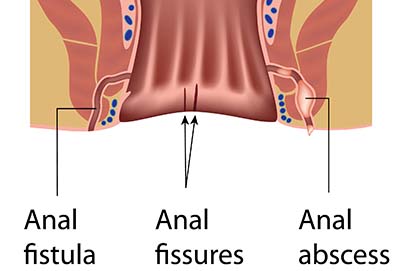
Anal Fistula

WHAT IS ANAL FISTULA?

Anal Fistula is caused by blokage of gland canals located in the anus, which soften and lubricate the stool during the toilet. The secretion channels blockage is due to different reasons probably development of infection. The developing infection either causes abscess formation in this region (anal abscess) or the existing abscess becomes chronic and manifests itself as a tunnel formation (anal fistula) between the anus canal and the skin. It is a disease that is difficult to follow and treat for both our patients and us. Because the area is dirty and open to infection.
Although it is more common in men, hormonal factors are held responsible for the development of the disease.
Said tunnel-shaped line (fistula tract) has two holes. The part defined as the inner mouth is the invisible starting point located in the breech canal, and the outer mouth is the end point located on the skin at the breech entrance and can be easily selected from the outside. Although the length of the tunnels can vary from 2 cm to 6-7 cm, it is the length of the existing line, its relationship with the breech muscles and its course that affect the course of the disease.
CAUSES
Usually the cause is an infection of the glands in the anal canal for any reason. Other causes can be listed as:
- Anal trauma (during accident, injury, birth trauma or enema application)
- Anal surgeries (interventions and surgical procedures due to hemorrhoids)
- Anal canal or rectum cancers (sometimes it can give symptoms in the form of fistula!!)
- Hard, indigestible ingredients in food (fish bone, egg shell)
- After radiotherapy applied to the last part of the intestine
- With inflammatory bowel diseases (chron and ulcerative colitis)
- It can be seen in people with diseases such as diabetes, HIV, tuberculosis, immune deficiency.
SYMPTOMS
Generally, patients’ complaints; a foul-smelling, yellow discharge from the anus and itching associated with it. It can be in the form of contamination in the underwear, or sometimes it is accompanied by bleeding. Hardening and skin discoloration due to chronic infection around the outer mouth, and a feeling of fullness in the rectum after defecation can be seen.
In case of severe pain, swelling and redness around the anus, it is considered to be an anal abscess. This situation is urgent and this area must be evacuated surgically.
HOW TO DIAGNOSE?
The patient’s history (current diseases, surgeries) is very important. Then, the diagnosis can usually be made by anal examination. The external mouth is investigated around the anus, after the external mouth is seen, rectal tapping (finger examination) should be made and the bulge line formed by the fistula on the anus wall should be felt by hand. When this line is rubbed, it can be seen that there is a yellow discharge from the outer mouth.
An anoscopic examination is required for the evaluation of the inner mouth, but we prefer imaging methods in order to reveal all aspects of the fistula line. In this sense, the most frequently applied method is Magnetic Resonance (MR) imaging. It is very important for us in terms of showing the presence of abscess in this region, the length of the fistula line, how many there are and its course (its relationship with the breech muscles). Sometimes, during MRI, imaging can be performed by administering drugs (contrast material) externally (MR fistulography).
Another method for determining the inner mouth is the examination performed under anesthesia. This examination, which is performed under general anesthesia, is for both diagnostic and therapeutic purposes, especially in patients with a complicated fistula.
TYPES OF ANAL FISTULA
Especially the course and placement of the line is very important in the surgical success of this disease. Because in this region, there are muscles that provide stool control we have to protect. Briefly, there are two muscle (sphincter) layers on the anus. Of these, the muscle located on the outside is the muscle that provides voluntary control (the muscle that allows holding the stool and relaxing and defecating in the toilet). There are the cases where the fistula line is associated with these muscles. This is the part that complicates the work. Because the type of surgery to be performed must be planned in advance and be specific to the patient and protect these muscles.
Anatomically, the types of anal fistula, according to the relationship with the muscles and the course it follows:
- The most common intersphincteric
- Second most common Transsphincteric suprasphincteric extrasphincteric
SURGERY DECISION
After the disease is diagnosed, anal fistula surgery should be planned as soon as possible. If the patient remains indecisive or delays the operation, it is possible for the fistula to spread and become complicated, making the treatment process more difficult. Even in patients who have complaints for many years and have not received treatment, serious pictures ranging from chronic irritation to anal cancers can be seen.
During periods when the discharge is very intense in fistula patients, antibiotic treatment is started at the first stage and the inflammation is expected to regress, and in this way, the patient is prepared for surgery. In the meantime, the decrease and disappearance of the discharge and the regression of the complaints do not mean that the disease is cured.
In patients with fistula that comes in the form of anal abscess, the abscess should be surgically evacuated under local anesthesia or general anesthesia, and then the inflammation should be removed with medical treatment (at least 2 weeks should be treated). ) will lay the groundwork.
SURGERIES FOR ANAL FISTULA
The gold standard in the treatment to be done is to eliminate the existing line and to preserve the muscle structures and not to deteriorate the stool control.
The method we frequently apply today is the FILAC (FistulaLaserClosure, laser closure) technique. The inner mouth is determined by entering the anal canal with a speculum under spinal or general anesthesia. Afterwards, the fistula line is closed by providing destruction through heat and light, thanks to the fiber with a laser at the end.
No incision during surgery
Short operation time
Absence of complications such as stool and gas incontinence after surgery
The patient can be discharged on the same day
Minimal post-operative pain
It is a safer option compared to classical methods, due to the short post-operative recovery time.
Another surgical method is fistulotomy. It is the method of opening the fistula line and leaving the body to its own healing process after cleaning the dirty area inside the tunnel. It is generally preferred in uncomplicated fistulas where the line is short.
Seton is a method used in fistulas containing a large amount of muscle tissue, where it is not possible to remove or cut the fistula line. It can be applied in two ways.
In loose seton application, a non-absorbable material is passed through the fistula line and left there. The aim is to ensure that the current flow and infection flow out of the material, as well as to close the line thanks to a controlled repair process called foreign body reaction.
In the cutting seton application, it is aimed to gradually cut the existing muscle tissue by squeezing the material used once a week. The disadvantage is that the tightening process is painful.
Complications such as temporary or permanent loss of stool and gas control and recurrence of the disease can be seen after both seton and fistulotomy techniques.,,